This month I have been teaching 1st year Lincoln Undergraduates Neuroscience for the first time. I started teaching this topic at Exeter University 10 years ago and over that time I have developed materials, resources and approaches which address some of the problems lecturers often face in teaching psychology students about the brain.
One problem is that neuroscience is often taught to students as “facts” rather than hard earned knowledge acquired from research studies, but unlike other areas of psychology it is difficult to find published studies in neuroscience suitable for using as the basis for a tutorial. Another problem is that very few psychology departments have facilities to give 200 undergraduates hands on experience of electrophysiological techniques or dissection of real brains, which limits what can be done in a lab class format.
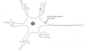
As a solution I devised several small group tutorials which engage the students in active problem solving tasks. In our “neuropsychology clinic” students are given patient case descriptions (based loosely on real cases). They work in groups and in turns request items of further information to view from the patients “records”, including various different neuropsychological test results, visual perimetry plots, MRI scans etc.. They have to use this information to work out a diagnosis e.g. apperceptive agnosia. In the “Neural Communication tutorial”, amongst other activities, students work through a series of cartoons depicting neuronal dendritic trees with several incoming axons and synapses, each with a different specified excitatory or inhibitory weight and action potential activity. Their task is to work out whether the neuron will reach a given threshold and “fire” or “not fire” its own action potential and the adding, subtraction and multiplication involved gets more and more complicated in each successive problem. This exercise helps students understand the work of John Eccles on post-synaptic potentials (PSPs), processes of temporal and spatial summation of PSPs and how computational processes occurring at the cellular level relate to the psychology of choice and decision making. 
Finally in our “Brain Lab practical” students visualise structures in the brain on a real 3D MRI scan. They learn how to operate a commonly used software tool for neuroimaging research in order to visualise cortical and sub-cortical brain structures. Students are then given a series of scans of real patient brains alongside a series of patient case descriptions and have to match the MRI scan to the correct case description (e.g. Brocas area stroke; fronto-temporal dementia). Brain models, atlases, text books, colouring books and brain hats! are made available in the classroom to add to the fun.
Both tutorials can be run by post-graduates with little advanced specialist knowledge of the topics covered, will work with groups of up to 25 and last about an hour each. The practical class requires MRIcron software to be installed (free) and fills a 2 hour session.
Please let me know if you are interested in using or adapting any of these resources in your course and I will be glad to send you the supporting materials.