Visual Attention and Paeleolithic Stone Tools

This month we published a study describing how people direct eye movements during viewing of ancient stone tools. The work was led by Maria Silva Gago, Emiliano Bruner and other researchers from CENIEH University of Burgos Spain together with myself and former Lincoln PhD student Flora Ioannidou (now at University of Aberdeen).

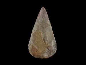
The research used eye tracking and the “mouse click” attention tracking technique to measure which areas of stone tools attracted peoples interest most. High resolution photographs of Paeleolithic hand axes (example right), along with much more ancient (approximate 2 million year old) roughly worked tools (example below) were shown to participants. We found that people’s attention was drawn to the “knapped” surfaces of  tools, as well as areas such as the base where the tool would normally be grasped. This was the case even though participants were just viewing pictures and not handling the objects and weren’t told the objects were tools.

We also ran the images through a computer model which calculated the”salience” of different regions of the images.  Some very influential models of vision suggest that attention is simply attracted to regions that stand out visually from the background (i.e. are more “salient”), but the computer model did not seem to explain our results very well. Instead, we think it was how the different parts of the tool might be grasped and used, rather than just how visually interesting it was, that was directing the viewers’ eyes.

The results support the theory of object-action affordances proposed by James J Gibson in the 1970s. He suggested that the brain very quickly detects features of objects that are relevant for action, priming a tendency for us to carry out the associated action. One possibility is that through evolution our ancestors’ brains became increasingly sophisticated at detecting action affordances in stone objects. This lead in turn to the manufacture of more sophisticated tools with enhanced features designed to activate action affordances. This in turn may have caused further development in the brain’s object-action affordance network in an ongoing process of coevolution between human cognitive capacities and tool complexity.

The full paper is published in the journal Perception and can be read online here.

Some example eye movement sequences recorded in the study are shown below.

Orienting to social cues in Parkinsons

This month we published a paper in the journal Experimental Brain Research reporting how eye movements are influenced by social cues in people with Parkinsons disease.

Previous studies have suggested that Parkinsons patients have problems in directing visual attention due to loss of the chemical dopamine within the basal ganglia of the brain, but no one has specifically looked at whether they have particular problems with social cues. This question is important as problems with processing social information may lead to difficulties in everyday life for people with Parkinsons. It is also a question of scientific interest as it has been suggested that we have specialised brain pathways for processing social information (the so-called “Social Brain hypothesis”).

We used a task in which pictures of someone else’s eyes, arrows and pointing fingers are shown on a computer screen, whilst people track a spot jumping around the screen with their eyes (see picture; you can see a video of the task here). Often people make an eye movement by mistake in the direction indicted by the eyes, arrows or fingers, even when they are meant to respond directly at the target (the little black spot) and ignore the pictures. We found healthy adults made more of these errors with pointing finger cues compared to the other cue types (suggesting pointing fingers provide a particularly strong cues to eye movements) whereas people with Parkinsons made similarly high errors for all 3 cue types.

Although not specifically better or worse for socially relevant cues (eyes / fingers), Parkinsons patients clearly had difficulty in suppressing their distracting influence. This suggests the basal ganglia plays a role in controlling eye movements in response to social and non-social cues. But this doesn’t mean that people with Parkinsons don’t have particular problems with maintaining attention in social situations. On the contrary, social interaction might often take place in a busy environment, with lots of sensory information competing for attention. These deficits in the control of eye movements might therefore lead to difficulty in social interaction and might impact on patients’ quality of life.

A link to the paper can be found here

Assessing premorbid IQ with picture vocabulary tests

An important task in neuropsychology is establishing how a patient’s current state compares to how they were before they suffered  brain injury. Assessing “premorbid” IQ is also of key importance in research studies that compare cognitive abilities in patients with a group of healthy participants, as it is important that the two comparison groups are as well matched as possible.

The most common way of assessing premorbid IQ to date is the National Adult Reading Test or “NART’. This short word reading test includes words with regular and irregular spellings and is highly correlated with IQ, whilst also “holding” after brain injury (being relatively unaffected relative to other tests of cognitive function). The problem with the NART is that it requires patients to read words out loud. Brain injury can also impair speech ability meaning that the test is impossible to use in patients with conditions such as aphasia.

We have recently published a paper in the journal Applied Neuropsychology: Adult which examined the effectiveness of the British Picture Vocabulary Scale (BPVS) as an assessment of premorbid ability. In this test patients’ must point to pictures matching the words spoken by the examiner. Originally developed to assess vocabulary in young children, we found that the BPVS appears to be at least as good as the NART at “holding” after brain injury and assessing premorbid IQ.

Along with other similar picture based tests, we think the BPVS could be a useful tool to assessing premorbid IQ in research as well as clinical neuropsychology practice.

Eyelander game evaluation and Parkinsons and Spatial Memory studies published

Research in patients both young and old can be difficult, time consuming and stressful to carry out (e.g. due to the ethical approval process, patient recruitment and practical difficulties in testing patients with physical disabilities etc). Yet the importance and potential benefits to patients themselves of such research far out weighs the difficulty entailed in conducting it.

Two of my recently published papers reflect the outcome of patient based projects. Both studies use tasks which require viewers to search through items on a screen using saccadic eye movements. The first addressed the issue of working memory and oculomotor control in Parkinsons disease, a topic I have been researching since the late 1990s. Whilst the second reports the clinical trial evaluating the effectiveness of the Eyelander video game for children who have had neurological injury leading to partial visual field loss (hemianopia).

In the first study, published in the April 2019 edition of the Journal of Cognitive Neuroscience we recorded eye movements while participants performed a version of the CANTAB Spatial Working Memory task which requires patients to search through boxes on a computer screen to find hidden tokens. I first had the idea to do this study whilst watching patients performing this task on a touch screen when I was a post-doctoral research fellow at Charing Cross Hospital, London. I could see that patients were using eye movements a lot in this token “foraging” task, but at the time we didnt have the technology to track their eye movements properly. It was only later that suitable eye tracking equipment and software became available to carry out the research. Amongst other findings the paper shows that people with Parkinsons don’t use eye movements to plan ahead or look back at locations they’ve already searched as effectively as controls, most likely due to an imbalance of the neurotransmitter dopamine in the prefrontal cerebral cortex.


The second paper, published in the December 2018 edition of Journal of Visual Impairment and Blindness describes the evaluation of our visual search  game for children with partial visual loss following brain injury affecting the visual parts of the cerebral cortex. The results showed children were able to play the game at home unsupervised and that it had a positive effect on parallel measures of functional visual ability which was similar in magnitude to effects reported for visual search training in adult with partial visual loss following stroke. The Eyelander game is now available for anyone to play online, so please take a look. We are also starting a collaborative project with Great Ormond Street Hospital to evaluate its effectiveness for treating visual field loss following neurosurgical procedures in children.

“Busy Bee” paper published in Experimental Brain Research

We have a paper published this month in the journal Experimental Brain Research based upon 2 years of data collected at the Lincoln Summer Scientist event which looked at how children’s saccadic eye movements are affected by directional socio-biological cues (see previous posts here and here).

We report results from 137 children who performed a pro-saccade task presented as  a computer game in which they had to keep their eyes on a cartoon bee that jumped unpredictably from the middle to the left or right of a computer screen. The Eyelink II system was used to examine how quickly and accurately the children followed the bee while pictures of arrows and photos of pointing hands and eyes appeared in middle the screen just before the “buzy bee” character moved (see Youtube video).busy bee task

We found that children were distracted by the direction of the pointing pictures such that their eyes were quicker to move towards the cartoon bee when she jumped in the same (Congruent) as opposed to the opposite (Incongruent) direction to the pointing finger, eye or arrow. Interestingly for the youngest group of children (3-5 years) this effect was found most strongly for pointing fingers. Only older children showed the effect for eyes and arrows. The paper makes the case for the view that children have to learn to link what they see in the world around them with the direction of interesting information and events. One of the first “cues” to attention that young children learn may be the direction of an adult’s pointing index finger.

Another interesting finding of the work was that for the youngest group of children when eye gaze cues overlapped with the onset of the peripheral target bee a large proportion of “omission errors” were made such that they missed the target completely and didn’t make a saccade. I was involved in the testing myself at Summer Scientist and I found this feature of young children’s behaviour particularly fascinating. It seems strikingly similar to stimulus “extinction” and ommission errorsneglect seen in adult stroke patients. Rather than just not moving their eyes I think 3-4 year olds didn’t “see” the Bee under these conditions and this is something I’d hope to follow up at future summer Scientist weeks.

The paper is full open access and available here . See here for another recent Experimental Brain Research study I’ve only recently seen by a group in Oslo showing ERP response to finger pointing cues in babies.