“Functional Pleomorphism” and the Neuroscience of Rules

Last month we published a paper in Frontiers of Human Neuroscience. This was the latest and last functional magnetic imaging (fMRI) study to come out of a Wellcome Trust funded project run by myself (whilst at University of Exeter) and Dr Ben Parris (now at Bournemouth), investigating the organisation and function of cognitive control processes in the human frontal cortex.pic2
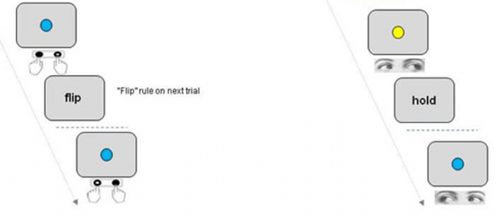
Humans are perhaps uniquely able to represent information in an abstract way which allows them to generalise rules and knowledge across different situations and tasks. For example we might learn to get a reward by pressing a button on the left with our finger when we see a blue shape, and a button on the right when we see a yellow shape. But we can also generalise rules to different situations e.g. instead of pressing a left or right button,make an eye movement to the left or right when we see the right colour…or say the word “left” or “right”…or wiggle the toes on your left and right feet!

This problem might seem trivial at first, but it is actually very difficult to see how neurons in the brain could achieve this, when ultimately they just make connections (“synapses”) linking inputs (a sensory stimulus) with outputs (a specific muscle movement). Imagine a neuron that truly represented the abstract concept of Blue things =Leftness. The only way this clever “concept neuron” could make us contract just our left interosseous muscle (the muscle in your index finger), or just our lateral extraocular muscle would be to have synapses that change their configuration completely within a fraction of a second so that they just sent signals down the connections to the finger or the eye muscles. Otherwise the signals from the neuron would either do nothing or have to make our whole body (including our toes!) move left. The idea that cells can change their synapses very rapidly (<1 second) is called neural functional pleomorphism and we don’t think it happens in the human brain.

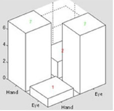
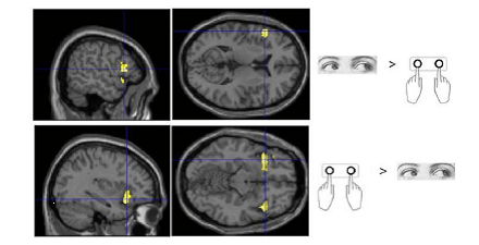
In our study we got participants to perform a Blue / Yellow colour – Left/Right response rule switching task in the brain scanner. Sometimes they had to make just an Eye movement and other times just a left/right button press. We found areas of the brain that responded just for Eyes and just for Hands as you might expect. But other areas in the prefrontal cerebral cortex didn’t care about the response and showed activity more related to the rules. But when we zoomed in on these same areas and analysed the pattern of activity at a finer scale during Hand and Eye “epochs”, there were significant differences between the two. So whats going on?

pic1


The answer, we think, is that there aren’t any “concept neurons” in your brain. Instead rules and concepts are embedded in the distributed pattern of activity across networks of neurons. Some of these individual cells might prefer eye movements to the right when we see a Blue Square, others may send signals to our left finger when we see a Blue Circle and others which make connections relating to all sorts of different versions and combinations of the general rule: blue things=left. When all the neurons representing the various different examples of the general rule work together, that is when we experience ourselves thinking about the general rule concept. But in order to actually do anything useful only the sub-set of neurons coding one specific “exemplar” of the rule is active.
pic3So there is no single place in our brain where task rules can be said to be represented and exist!

The actual paper is less philosophical but it is Open Access and you can download it here
Quality Adjusted Life Years and the Neuroscience of Fairness

This month we published the first ever fMRI brain imaging study of health care rationing decision making 

pic1
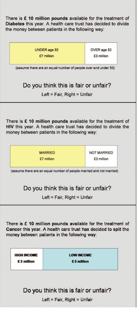
decision making screens

Health care funders around the world have to make difficult moral decisions to allocate limited money to treat various medical conditions in different groups of patients. For example whether or not to support funding for expensive new drug treatments for a group of cancer patients with low chance of survival relative to a drug that will benefit a group with a higher chance of survival.  A commonly used framework on which these decisions can be based are Quality Adjusted Life Years or “QALYs”. This system is based on so-called utilitarian decision making principles, which prioritize choices that deliver the maximum benefit to the greatest number of people. The problem with QALYs  is that decisions based on this approach are often viewed negatively by members of the general public. People instead believe that everybody has a “right” to receive medical care and anything that violates this principle is unfair and immoral.

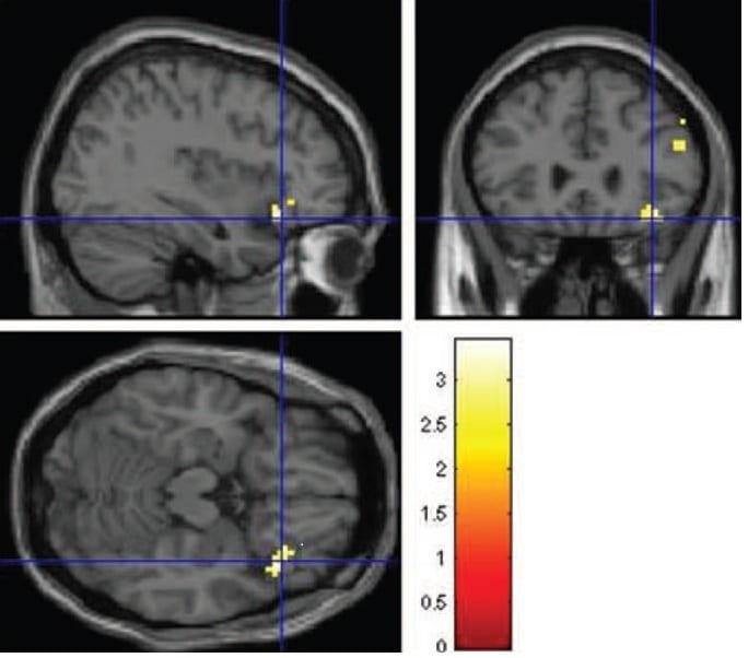
Consistent with previous work, when asked to judge the “fairness” of various scenarios depicting a split between different social groups our participants judged unequal division of funding as “unfair”, even when principles of QALY might indicate otherwise. Interestingly brain regions linked to emotion as well as cognitive processes were active during decision making. Unequal splits of resources for medical care produced activity in the anterior insula, a region often associated with social / moral disgust (see earlier posts on social norms and eating disorders). Further, under conditions where participants were prepared to judge unequal splits as fair, more activity was seen in the inferior frontal cortex, a region activated when humans inhibit a strong response impulse.

The results represent a preliminary first step for cognitive neuroscience into the field of health economics and the paper is careful to avoid over interpreting the findings and applying them to real situations outside the scanner. But the findings are consistent with a bigger idea that humans have two decision making systems, one cognitive and one more emotional / instinctive. Given enough information people may be more inclined to support healthcare decisions based on QALYs, but this requires cognitive effort to over-ride a more emotion based bias towards absolute equality and universal rights.

The paper is out  ipic2n the June edition of the Journal of Neuroscience Psychology and Economics. The research was carried out in collaboration with Prof. Paul Anand ( Open University and Health Economics Research Centre at the University of Oxford), Lisa Smith (Flinders University Austrailia) and the Exeter Magnetic Resonance Research Centre. A pre-print of the paper is available via the Lincoln Repository website.
Award for Journal of Neuroscience Psychology and Economics paper

Our 2012 fMRI “Neuro-economics” study of a Coordination / Temptation game scenario, carried out in collaboration with Milan based social philosopher Francesco Guala has been given an award as one of the Top 10 all time classic papers published in the APA publication JNPE. I am looking forward to traveling to Munich to accept the award at the NeuroPsychoEconomics conference at Ludwig-Maximillian University at the end of May.

Abstract of paper

More about the conference 

Psychology News Blog 

And previous posts

MRI-scan-of-human-brain-008

Learning stimulus-saccade mappings in Parkinsons

In our most recent research paper (published this month in the journal Neuropsychologia ), we investigated how people with Parkinsons perform a computerised eye movement rule switching task, which we have used previously in patients with frontal lobe damage (see U-tube video: and Summer Scientist 2012).

Many every day tasks require us to learn to make links between what we see and where we look with our eyes. We also need to be able to switch between performing one task or another (e.g. making a cup of tea, reading the newspaper and then answering the telephone) and learn new skills such as preparing a new recipe or learning a new game or sport.

Unlike patients with frontal lobe strokes, People with Parkinsons didn’t show any big problems in switching between stimulus-saccade “rules” (e.g. blue stimulus = look left), but were slower to learn a new rule by trial and error learning compared to participants without Parkinson. This suggests that the brain circuits and chemicals affected in Parkinsons play a role in this ability and that people with Parkinsons may have problems learning new visuo-spatial tasks over and above the obvious difficulties the condition causes with movement.

Parkinson's disease

Please contact me if you would like a reprint of the paper or would like to know more about this research.


Post hypnotic suggestion and the Stroop effect

Over the last two decades the use of hypnosis and post-hypnotic suggestion has moved from the realm of parapsychology into the mainstream of cognitive neuroscience research into consciousness. Former Wellcome Trust funded post-doc Ben Parris (University of Bournemouth) has developed a particular interest in how “cognitive control” mechanisms can be modified by post-hypnotic suggestion. The process of inducing an hyponotic suggestion in the lab involves reading out a simple script to volunteers who have consented to be hypnotised and have been identified to be susceptible to hypnosis through a pre-screening procedure. 

  Our most recent paper together with Zoltan Dienes (Universityof Sussex), just published in the Journal of Experimental Psychology: Human Perception and Performance, examined how hypnotic suggestion affected performance in the Stroop task. In the classic Stroop task participants have to respond to the ink colour of a word which can be in conflict with the word itself e.g BLUE. Normally people respond more slowly when the word and colour are in conflict with each other, but previous studies have suggested that the effect can be abolished by implanting the suggestion that words are printed in incomprehensible characters from a foreign language (the “word blindness” suggestion)(Raz et al. 2002).

  Ben was only partially able to replicate the original results and showed that it was only under certain conditions that the Stroop effect was abolished. Specifically when the stimuli were presented rapidly such that there was only 500ms between the last response and the next word being shown. Subjects who did not receive the word blindness suggestion still showed a strong Stroop effect even at the short response stimulus intervals.

   The finding tells us about the level at which the hypnosis effect influences control over mental processes. In the case of the Stroop effect, word blindness is “activated” in a reactive way by the onset of a word and participants do not always exert sustained effort to maintain the word blindness suggestion. How post-hypnotic suggestions affect the brain’s perceptual and action control centres remains an ongoing question, but together with previous work, the new results offer some intriguing clues. They also confirm that hypnotic suggestion is a serious tool for the scientific study of mental processes and consciousness.

References/links

Parris BA, Dienes Z, Hodgson TL (2012) Temporal Constraints of the Word Blindess Posthypnotic Suggestion on Stroop Task Performance. Journal of Experimental Psychology: Human Perception and Performance. Advance online publication. http://psycnet.apa.org/psycinfo/2012-10032-001/

Raz A et al. (2002) Suggestion reduces the Stroop effect. Psychological Science, 17, 91-95.

OAKLEY, D. A. & HALLIGAN, P. W. (2009). Hypnotic suggestion and cognitive neuroscience. Trends in Cognitive Sciences, 13, 6, 264- 270  http://huuk.nfshost.com/