Last month we published a paper in Frontiers of Human Neuroscience. This was the latest and last functional magnetic imaging (fMRI) study to come out of a Wellcome Trust funded project run by myself (whilst at University of Exeter) and Dr Ben Parris (now at Bournemouth), investigating the organisation and function of cognitive control processes in the human frontal cortex.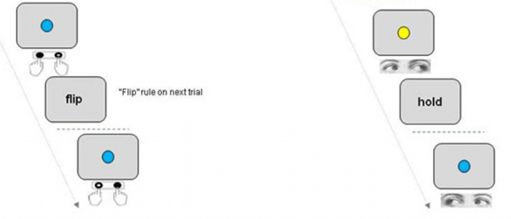
Humans are perhaps uniquely able to represent information in an abstract way which allows them to generalise rules and knowledge across different situations and tasks. For example we might learn to get a reward by pressing a button on the left with our finger when we see a blue shape, and a button on the right when we see a yellow shape. But we can also generalise rules to different situations e.g. instead of pressing a left or right button,make an eye movement to the left or right when we see the right colour…or say the word “left” or “right”…or wiggle the toes on your left and right feet!
This problem might seem trivial at first, but it is actually very difficult to see how neurons in the brain could achieve this, when ultimately they just make connections (“synapses”) linking inputs (a sensory stimulus) with outputs (a specific muscle movement). Imagine a neuron that truly represented the abstract concept of Blue things =Leftness. The only way this clever “concept neuron” could make us contract just our left interosseous muscle (the muscle in your index finger), or just our lateral extraocular muscle would be to have synapses that change their configuration completely within a fraction of a second so that they just sent signals down the connections to the finger or the eye muscles. Otherwise the signals from the neuron would either do nothing or have to make our whole body (including our toes!) move left. The idea that cells can change their synapses very rapidly (<1 second) is called neural functional pleomorphism and we don’t think it happens in the human brain.
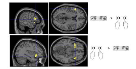
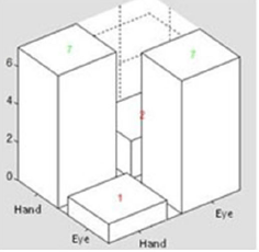
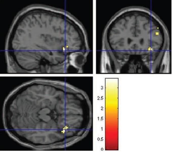
In our study we got participants to perform a Blue / Yellow colour – Left/Right response rule switching task in the brain scanner. Sometimes they had to make just an Eye movement and other times just a left/right button press. We found areas of the brain that responded just for Eyes and just for Hands as you might expect. But other areas in the prefrontal cerebral cortex didn’t care about the response and showed activity more related to the rules. But when we zoomed in on these same areas and analysed the pattern of activity at a finer scale during Hand and Eye “epochs”, there were significant differences between the two. So whats going on?
The answer, we think, is that there aren’t any “concept neurons” in your brain. Instead rules and concepts are embedded in the distributed pattern of activity across networks of neurons. Some of these individual cells might prefer eye movements to the right when we see a Blue Square, others may send signals to our left finger when we see a Blue Circle and others which make connections relating to all sorts of different versions and combinations of the general rule: blue things=left. When all the neurons representing the various different examples of the general rule work together, that is when we experience ourselves thinking about the general rule concept. But in order to actually do anything useful only the sub-set of neurons coding one specific “exemplar” of the rule is active.
 So there is no single place in our brain where task rules can be said to be represented and exist!
The actual paper is less philosophical but it is Open Access and you can download it here
 Our research has shown that there is a particular “marker” of Parkinsons in eye movements, namely “multi-stepping” or jerkiness of eye movements measurable under certain conditions with a computerised eye tracker. In other situations some people with Parkinsons appear to be slightly more distractable and not organise eye movements as efficiently as healthy people when carrying out problem solving and memory tasks.


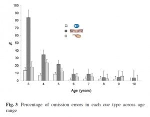 neglect seen in adult stroke patients. Rather than just not moving their eyes I think 3-4 year olds didn’t “see” the Bee under these conditions and this is something I’d hope to follow up at future summer Scientist weeks.
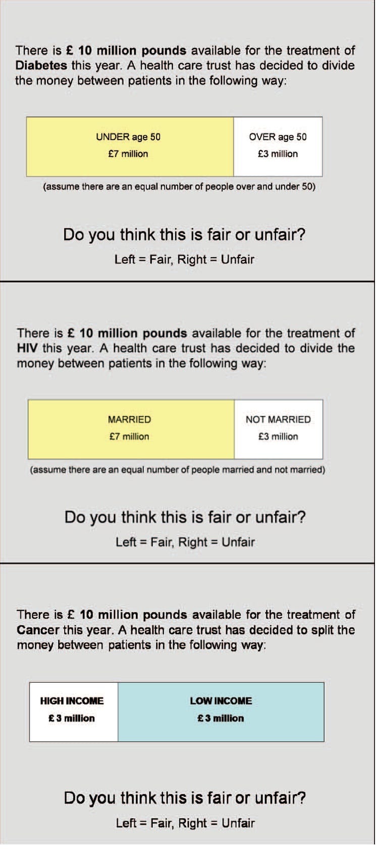
 n the June edition of the Journal of Neuroscience Psychology and Economics. The research was carried out in collaboration with Prof. Paul Anand ( Open University and Health Economics Research Centre at the University of Oxford), Lisa Smith (Flinders University Austrailia) and the Exeter Magnetic Resonance Research Centre. A pre-print of the paper is available via the Lincoln Repository