The fingers, not the eyes, have it

Next week we will be presenting our work on orienting to socio-biological cues in children at the 17th European Conference on Eye Movements in Lund, Sweden.

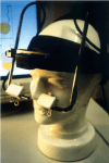
me and eyetrackerAt last years Lincoln Summer Scientist event we asked children to play a game in which they followed a cartoon bee jumping to the left or right with their eyes whilst distracting pictures of arrows, pointing fingers and someone else’s eyes gazing to the left or right were shown in the middle of the screen.  We tracked their eye movements with an Eyelink 1000 eye tracking system and measured how quickly they made saccades to follow the bee.

We found that the youngest children (4-5 year olds) showed a large “congruency” effect for pictures of pointing fingers such that their speed of looking was slowest when the bee jumped in the opposite direction to that in which the hand pointed. Surprisingly, although the pre-schoolers weren’t similarly affected by pictures of eyes and arrows, older fellow summer scientists showed an equally strong congruency effect for all three types of cue (hand, eye and arrows).

The results are potentially very interesting and important in respect to understanding the best ways to direct young children’s attention quickly and effectively in an educational context as well as keeping them away from harm inside or outside of the home, but they might also have more profound implications. Rather than having hard wired “social brain” systems for processing socio-biological stimuli as suggested by some theorists, instead the brain may learn to form fast connections between what we see and what we do in early childhood. It just happens that pointing fingers may be among the first cues children learn to use in this way.

busy bee

We’re looking forward to finding out what other researchers think of our results in Lund and plan to replicate the finding at this years Lincoln Summer Scientists event. The work is carried out in collaboration with Nicola Gregory (Bournemouth University Face Research Centre). The work is part supported by the WESC foundation for Childhood Visual Impairment.

Slide1


Inaugural Lecture: Tuesday 9th April 6pm

Next Tuesday I will be giving my public inaugural lecture at the University of Lincoln. I have chosen to focus on ways in which human saccadic eye movements have and may in the future have an impact beyond academia. I will explain how eye movements can change the world in 4 different ways:

1. Eye movements themselves change our perception of the world. In a sense we all see the world differently in a way affected by our individual eye movements;

2. Visual search training, improving the efficiency with which we use eye movements, can be used to improve functional vision in adults and children with visual problems;

3. Research into attention and eye movements might be brought to bear to improve navigational cues in our visual environment, such as road signage;

4. Finally and perhaps most exciting is that saccadic measures might not only be valuable in the assessment of patients with neurological disorders, but could potentially be used to provide an early diagnosis of such conditions as Parkinsons and Dementia.

eye

   I am looking forward to talking to a general audience including members of the public, colleagues, friends and family and hope they will agree that eye movements can indeed change the world!

More details of the lecture and how to book are given here:

http://www.lincoln.ac.uk/home/campuslife/whatson/eventsconferences/event,name,20188,en.html

KTP Grant Success – Improving functional vision in children through a visual search computer game

We have been awarded a Knowledge Transfer Partnership (KTP) grant worth approximately £130K to support an exciting project which aims to apply visual neuroscience to the rehabilitation of childhood cerebral visual impairment and special education. The work is a collaboration between the University of Lincoln, Schoolof Psychologyand the West of England School and College for young people with little or no sight (WESC) (www.westengland.ac.uk). The grant will employ an experienced neuroscience / psychology researcher at WESC inExeter who will develop and evaluate a visual search rehabilitation computer game for use in children with partial visual loss. Dr Conor Linehan from the Lincoln Social Computing Research Centre (an expert in educational games) will also play a leading role in the project (http://staff.lincoln.ac.uk/clinehan).

WESC approached us as they realised that many of the children and young people they work with have problems which are due to damage to the brains visual centres rather than disorders of the eye itself. The project will also help WESC build expertise and understanding of the role of the brain in visual perception and its disorders. Previous research has demonstrated that visual search training can lead to significant recovery of vision following damage to visual regions of the brain in adults, but adult training programmes are simply to boring to use with children. At the same time, we expect that implementing visual search training as a game could also lead to improvements to provision of search training to adults with hemianopia (visual loss following stroke).

KTPs are a national initiative which supports partnerships between business and universities enabling Associates to work on challenging, high profile projects (www.ktponline.org.uk). Financial support for the KTP project with WESC is provided by the Technology Strategy Board with offer of a part contribution from the Medical Research Council.

Please let me know if you are interested in finding out more about this project. If you think you have a suitable background and are interested in applying for the position please see the job and application procedure here: http://jobs.lincoln.ac.uk/vacancy.aspx?ref=EL1076A

See also previous post: https://hodgson.blogs.lincoln.ac.uk/2012/05/28/visual-neuroscience-and-specialist-education/

Tutorials and practicals for biological psychology teaching

This month I have been teaching 1st year Lincoln Undergraduates Neuroscience for the first time. I started teaching this topic at Exeter University 10 years ago and over that time I have developed materials, resources and approaches which address some of the problems lecturers often face in teaching psychology students about the brain.

   One problem is that neuroscience is often taught to students as “facts” rather than hard earned knowledge acquired from research studies, but unlike other areas of psychology it is difficult to find published studies in neuroscience suitable for using as the basis for a tutorial. Another problem is that very few psychology departments have facilities to give 200 undergraduates hands on experience of electrophysiological techniques or dissection of real brains, which limits what can be done in a lab class format.

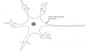
   As a solution I devised several small group tutorials which engage the students in active problem solving tasks. In our “neuropsychology clinic” students are given patient case descriptions (based loosely on real cases). They work in groups and in turns request items of further information to view from the patients “records”, including various different neuropsychological test results, visual perimetry plots, MRI scans etc.. They have to use this information to work out a diagnosis e.g. apperceptive agnosia. In the “Neural Communication tutorial”, amongst other activities, students work through a series of cartoons depicting neuronal dendritic trees with several incoming axons and synapses, each with a different specified excitatory or inhibitory weight and action potential activity. Their task is to work out whether the neuron will reach a given threshold and “fire” or “not fire” its own action potential and the adding, subtraction and multiplication involved gets more and more complicated in each successive problem. This exercise helps students understand the work of John Eccles on post-synaptic potentials (PSPs), processes of temporal and spatial summation of PSPs and how computational processes occurring at the cellular level relate to the psychology of choice and decision making.

   Finally in our “Brain Lab practical” students visualise structures in the brain on a real 3D MRI scan. They learn how to operate a commonly used software tool for neuroimaging research in order to visualise cortical and sub-cortical brain structures. Students are then given a series of scans of real patient brains alongside a series of patient case descriptions and have to match the MRI scan to the correct case description (e.g.  Brocas area stroke; fronto-temporal dementia). Brain models, atlases, text books, colouring books and brain hats! are made available in the classroom to add to the fun.

   Both tutorials can be run by post-graduates with little advanced specialist knowledge of the topics covered, will work with groups of up to 25 and last about an hour each. The practical class requires MRIcron software to be installed (free) and fills a 2 hour session.

Please let me know if you are interested in using or adapting any of these resources in your course and I will be glad to send you the supporting materials.

Research explores rule switching across the life span

You are an astronaut trying to boldy go where nobody has gone before… but two naughty aliens have stolen vital bits of your rocket motor, leaving you stranded on an asteroid in a very boring corner of the solar system. Luckily the aliens are simple creatures who like to use the same craters to hide their booty:  the colour of the alien tells you which one of the two craters (left or right) to look in. Your task is to work out which colour alien goes with which crater, but watch out as the tricky aliens keep swapping which crater goes with each colour!

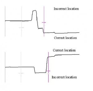
 How do our brains learn rules linking what we see with where we direct action? This simple ability is important for many visual tasks ranging from crossing the road safely to playing chess. Research from myself and collaborators has used an eye movement “Rule switching” task in which people have to learn rules linking a coloured shape (displayed on a computer screen) and a saccade (eye movement) to the left or right to get a “reward” (a smiley emoticon). We have shown that adult patients with different types of neurological and psychiatric problems have difficulty with this task. For example, damage to the frontal parts of the brain can lead to problems in switching between rules and stopping oneself making eye movements based upon the old rule.

At  the Lincoln summer scientist event this year we’ll be looking at how children of different ages do this task in order to gain insights into how rule learning and cognitive flexibility develops over the lifespan. For example, do children (whose brains’ haven’t finished developing yet) show any similarities to adults who have been unfortunate enough to have suffered strokes? Or, perhaps they might actually be better than older healthy participants who usually show a big performance “cost” when rules change and make more “corrective” saccades as if the old rule is still “active” somewhere in the brain.

  Of course, we’ve had to make the task a bit more fun for the young scientists who’ll be helping us with our research (hence the aliens and asteroids). We are also using our brand new Eyelink 1000 eye tracker for the first time as it allows “head free”eye tracking, making it much better for use with kids (see eye movements on U-tube).Visiting research student Rebecca Facey from the University of Exeter will also be helping out with the project.

It promises to be a busy but fun week, so its: 5..4..3..2..1 blastoff! forLincoln summer scientist 2012.

References

Huddy VC, Hodgson TL, Ron MA, Barnes TRE, Joyce EM (2011) Abnormal negative feedback processing in first episode schizophrenia: evidence from an oculomotor rule switching task. Psychological Medicine 41(9),   p 1805-1814.

Hodgson TL, Chamberlain M, Parris BA, James M, Gutowski NJ, Husain M, and Kennard C. (2007) The role of the ventrolateral frontal cortex in inhibitory oculomotor control. Brain, 130: 1525-1537.

Husain M, Parton A, Hodgson T, Mort D & Rees G (2003) “Self-control during response conflict by human supplementary eye field.” Nature Neuroscience. 6(2): 117-118.